Abstract The first step in the interpretation of mass spectra is always determination of the molecular weight. Then, the interpretation of observed the observed fragment ions and subsequent MS/MS experiments (if available) can help to determine the connectivity of the atoms. Exact mass determination with the mass accuracy better than 5 ppm allows assignment of the probable elemental formula fragment ions in the spectra. The last interpretation step is the proposal of a structure, which should be verified with measurement of authentic standard if available.
KeywordsInterpretation, Electron ionization, Soft ionization techniques, Molecular weight, Fragmentation, Functional group, Biomolecule
LevelBasic
For many decades, EI was the only ionization technique in mass spectrometry. As a result, the “rules” for the fragmentation and spectra interpretation are best explained for EI. Many of these rules transfer to soft ionization techniques as well. It should be noted that the rules for mass spectra interpretation are fraught with exceptions, which complicates spectral interpretation and tests the skill and experience of the researcher. Some useful rules and hints are listed here.
![]() Nitrogen rule
Nitrogen rule
For compounds composed of common organic elements (C, H, O, N, S, Si, P, F, Cl, Br, I),
- An odd MW automatically means an odd number of nitrogen atoms in the structure.
- An even MW indicates an even number of nitrogen atoms in the ion. (Remember that zero is an even number!)
This rule is valid for all neutral species (e.g. molecular weight determination) and odd-electron ions (OE+.).
For even-electron ions (EE+), the rule is just the opposite. In general, the nitrogen rule is applicable for all atoms having either:
- Even valence and even atomic mass (e.g. carbon, oxygen, sulfur, etc.) or
- Odd valence and odd atomic mass (e.g. hydrogen, chlorine, bromine).
| Nitrogen number | Odd m/z value | Even m/z value |
| Even and zero (0, 2, 4, ...) | EE+ | OE+. |
| Odd (1, 3, 5, ...) | OE+. | EE+ |
Even-electron rule
The fragmentation of an odd-electron ion may provide either:
- an odd-electron fragment ion and a neutral loss, or
- even-electron fragment ion and loss of a radical.
The Even-Electron rule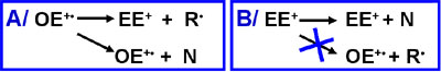 On the other hand, an even-electron ion may only provide even-electron fragment ions and the neutral loss, but not odd-electron ions and the radical loss. Some exceptions exist, however.
Isotopic abundances
Common organic elements can be divided into 3 groups according to the presence of their naturally occurring isotopes M+2 and M+1:
- M+2 elements (bromine, chlorine, sulfur, silicon and oxygen),
- M+1 elements (carbon and nitrogen)
- M elements (fluorine, iodine, phosphorus and hydrogen)
The natural relative abundance of deuterium (2H) is negligible (0.015%), hence hydrogen is included in the group of monoisotopic (M) elements.
Natural abundances of common organic elements
(Click here to view a more informative coloured version of this table)
| Element | "M" isotope | "M+1" isotope | "M+2" isotope | Type of element | |||
| m/z | % | m/z | % | m/z | % | ||
| H | 1 | 100 | 2 | 0.015 | - | - | "M" |
| C | 12 | 100 | 13 | 1.1 | - | - | "M+1" |
| N | 14 | 100 | 15 | 0.37 | - | - | "M+1" |
| O | 16 | 100 | 17 | 0.04 | 18 | 0.2 | "M+2" |
| F | 19 | 100 | - | - | - | - | "M" |
| Si | 28 | 100 | 29 | 5.1 | 30 | 3.3 | "M+2" |
| P | 31 | 100 | - | - | - | - | "M" |
| S | 32 | 100 | 33 | 0.79 | 34 | 4.3 | "M+2" |
| Cl | 35 | 100 | - | - | 37 | 32 | "M+2" |
| Br | 79 | 100 | - | - | 81 | 97.3 | "M+2" |
| I | 127 | 100 | - | - | - | - | "M" |
α-cleavage
Alpha cleavage is initiated by a radical center. The driving force is the formation of an electron pair from initial non-paired single electron, the preference order:
nitrogen > sulfur, oxygen, Π-electron, alkyl > chlorine > bromine > hydrogen
I-cleavage
Inductive cleavage is initiated by a positive charge center. The driving force is the attraction of an electron pair by the positive charge, the order of preference is: halogen > oxygen, sulfur >> nitrogen, carbon.
Molecular weight (MW) determination is always the first step in the interpretation of a mass spectrum. When the MW is not assigned correctly, all subsequent interpretation is useless. The figure shows a typical and trivial example of MW determination for a hypothetic compound with MW=500 Da.
Molecular weight determination (Click to enlarge)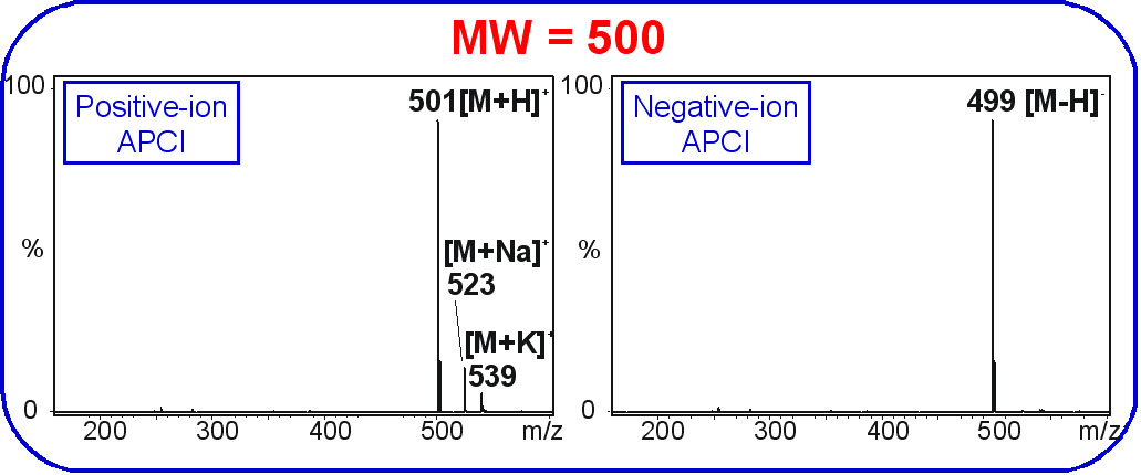
In the positive-ion mode, the base peak is the [M+H]+ ion, usually accompanied by less abundant peaks from adducts with sodium ion [M+Na]+ and ![]() potassium ion [M+K]+.
potassium ion [M+K]+.
The presence of peaks corresponding to low abundance alkali metal adducts may be considered proof of a correct MW determination because they are generally found only as molecular adducts, but there are exceptions: In negative-ion mode, the typical base peak is [M-H]- ion confirming the MW assignment.
Depending on the mobile phase composition, the salt content in particular, other adducts may be found, including [M+NH4]+ if the ammonium ion is present, adducts with solvents [M+H+methanol]+ or [M+H+acetonitrile]+, or even dimeric ions [2M+H]+, [2M+Na]+, etc.
As a rule of thumb, the base peaks should be [M+H]+ and [M-H]- in positive-ion or negative-ion mode, respectively, and the relative abundances of other adduct, dimers or fragment ions should have low to negligible relative abundances. Unfortunately, there are many specific cases where the appearance of full scan mass spectrum for soft ionization techniques may differ from this oversimplified ![]() scheme. The known exceptions not exhibiting (de)protonated molecules are:
scheme. The known exceptions not exhibiting (de)protonated molecules are:
- some organometallic compounds,
- polynitrated aliphatic compounds (i.e. explosives),
- some polysulfated compounds, etc.
There are also exceptions where radical odd-electron ions are formed instead of or in addition to common even-electron ions [M-H]- in the negative-ion mode. Examples include:
- (poly)nitro compounds known for the presence of M—. formed by electron capture in the negative-ion mode,
- highly conjugated polyaromatic compounds (especially those with heteroatoms),
- some organometallic compounds and some compounds containing multiple different functional groups.
Distinguishing odd-electron and even-electron ions.
For mass spectral interpretation, it is essential to distinguish between odd-electron (OE+.) and even-electron (EE+) ions.
All neutral molecules containing common organic elements (C, H, O, N, S, Si, P, F, Cl, Br, I) have an even number of electrons. When the molecule is ionized:
- by EI, one electron is lost, converting the species into an
 odd-electron ion (M+.)
 - by a soft ionization mechanism in the positive-ion mode, one proton (i.e. species without any electron) is added to the molecule, which causes no change in the number of electrons, which remains even, i.e. the even-electron ion [M+H]+ is formed.
The formation of ![]() even-electron [M-H]- ion in the negative-ion mode can be explained by analogy:
even-electron [M-H]- ion in the negative-ion mode can be explained by analogy:
- When a neutral non-radical species is lost during the fragmentation process (called "neutral loss") from any ion, then the number of electrons (either even or odd) remains unchanged.
- When a radical fragment is cleaved, then even/odd electron character is altered.
Functional groups have a significant influence on the ionization and fragmentation paths of individual compounds. The most suitable ionization technique and polarity mode may also be ![]() predicted by considering the functional groups present in the chemical structure.
predicted by considering the functional groups present in the chemical structure.
As a rough rule, the influence of functional groups on fragmentation behaviour follows the series:
sulfate > sulfonic acid > carboxylic acid > hydroxyl group > nitro group > halogens > other oxygen containing functional groups, etc.
Of course, this order is not strict and cannot reflect all combinations of elements, because the fragmentation process for compounds containing multiple functional groups may be rather complex with parallel ![]() fragmentation paths of varying importance.
fragmentation paths of varying importance.
One easy-to-understand mass spectrum demonstrating the correlation between observed fragment ions and the multiple functional groups is shown in the spectrum of sulfonated azo dye Mordant Black 15 measured in the negative-ion ESI-MS/MS mode:
Fragmentation scheme of Mordant Black (Click picture to enlarge)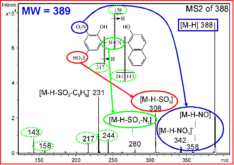
Examples of fragmentation ( Click to view all 5 schemes of fragmentation of the following functional ![]() groups):
groups):
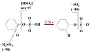
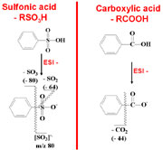
- The presence of the sulfate group is usually indicated by extensive fragmentation. The relative abundance of the deprotonated molecule [M-H]- can be significantly reduced in the full scan mass spectra. In the case of polysulfated compounds, the deprotonated molecule is negligible because of the consecutive losses of sulfuric acids [M-H-H2SO4]-, [M-H-2H2SO4]-, etc. The typical neutral losses are H2SO4 (Δm/z 98), SO3(Δm/z 80) and the typical low-mass diagnostic ion is [HSO4]- at m/z 97.
- The base peak in negative-ion spectra of monosulfonic acids is the [M-H]- ion, for polysulfonic acids a series of deprotonated molecules can be expected, such as [M-2H]2-, [M-3H]2- and in general [M-xH]x-.
- The typical feature of the mass spectra of sulfated and sulfonated compounds is the formation of sodium adducts such as [M-2H+Na]-, [M-3H+2Na]-, and in general [M-(x+y)H+yNa]x- ions. The highest observed charge, x, and/or highest number of protons replaceable by sodium ions corresponds to the total number of acidic groups in the molecule. The typical neutral losses observed in tandem mass spectra of sulfonic and polysulfonic acids are SO3 (Δm/z 80) and SO2 (Δm/z 64). The diagnostic ion is the radical-ion [SO3]-. at m/z 80. Obviously, the negative-ion mode is the method of choice for sulfates, sulfonic and carboxylic acids due to the ready formation of deprotonated molecules. Generally, positive-ion mode does not provide any signal for sulfates and sulfonic acids, especially when more than one functional group is present.
- A similar situation is found for carboxylic acids, though mono- and dicarboxylic acids containing other functional groups (e.g. hydroxyl or amino groups) can be also detected in the positive-ion mode, thought negative ion detection would be more sensitive for them. The base peak of negative-ion ESI or APCI spectra of carboxylic acids is [M-H]-. The characteristic fragment ion is [M-H-CO2]- ion (Δm/z 44) and is typically observed even in the full scan spectra. Generally, this neutral loss is clear evidence for the presence of carboxylic group. The hydroxyl group leads to an abundant peak corresponding to the neutral loss of water (Δm/z 18). The neutral loss of H2O does not prove the presence of the hydroxyl group because this neutral loss may be also observed for other oxygen containing functional groups, though usually with lower relative abundance and not in full scan spectra unlike alcohols/phenols.
- Alcohols and phenols can be analyzed in both polarity modes depending on the other functionalities in the molecular structure. The nitro group exhibits three potential neutral losses, NO2 (Δm/z 46), NO (Δm/z 30) and O (Δm/z 16). The last one is rather unusual (known only for N-oxides and some nitroso compounds) with very low to negligible relative abundance. The losses of NO2 and NO, however, are characteristic for the nitro group.
- When other more polar groups (sulfate, sulfonic and carboxylic acids) are absent, the presence of nitro group may cause the formation of the radical-ion M—. in negative-ion APCI mode as a result of electron capture, which is not too common with soft ionization techniques. Typically, the deprotonated molecule [M-H]- is observed in negative-ion mode with soft ionization techniques.
- The common neutral loss for compounds containing halogen atoms is the loss of HX. If multiple halogen atoms are present in the structure, consecutive losses are observed. Chlorine and bromine atoms show characteristic M : M+2 isotopic abundance ratios, approximately 3 : 1 for 35Cl : 37Cl and 1 : 1 for 79Br : 81Br.
Examples of fragmentation of functional groups:
(Click this text to view full scheme in pdf of sulphonate, sulphonic acid, carboxylic acids, nitro and azo-compounds, halogens, chlorine, bromine, chinones and phenols.)
For detailed studies of fragmentation mechanisms or quantitation, the analyte’s analogues labeled with stable isotopes in well-defined positions are used for comparison with non-labeled ![]() standards.
standards.
Two soft ionization techniques, ESI and MALDI, dominate the analysis of biomolecules. The full scan spectra usually provide (de)protonated molecules and molecular adducts useful for MW determination, while tandem mass spectra provide information on the sequence of particular building blocks in the structure, for example amino acids in peptides and proteins.
The established convention uses the assignment of a, b, c for fragment ions starting from N-terminus (amino group) and x, y, z for fragment ions from C-terminus (carboxylic group). The subscript is the number of amino acid as counted from the corresponding terminus of the polymer.
Peptide fragment nomenclature (Click to enlarge)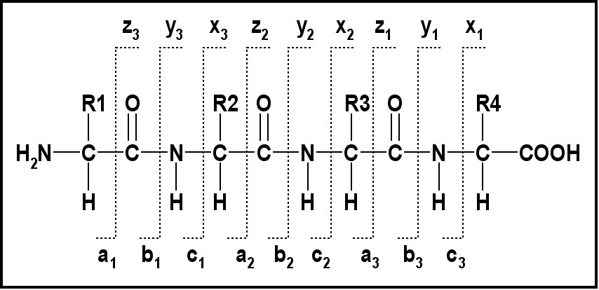
Spectrum of a protein Lysozyme, as an example of protein molecular weight determination:
ESI spectrum of Lysozyme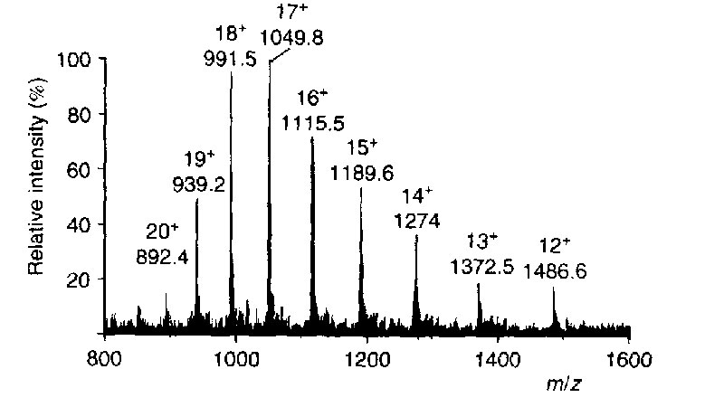
Unlike to most other compounds, the spectra of biomacromolecules can be found in extensive internet databases (for free or as subscription services), which significantly speeds up the identification of biomolecules previously measured. For "de novo" sequencing, the interpretation has to be done manually.